Hidden History

The Underdog Doctors Who Saved America’s “Blue Babies”

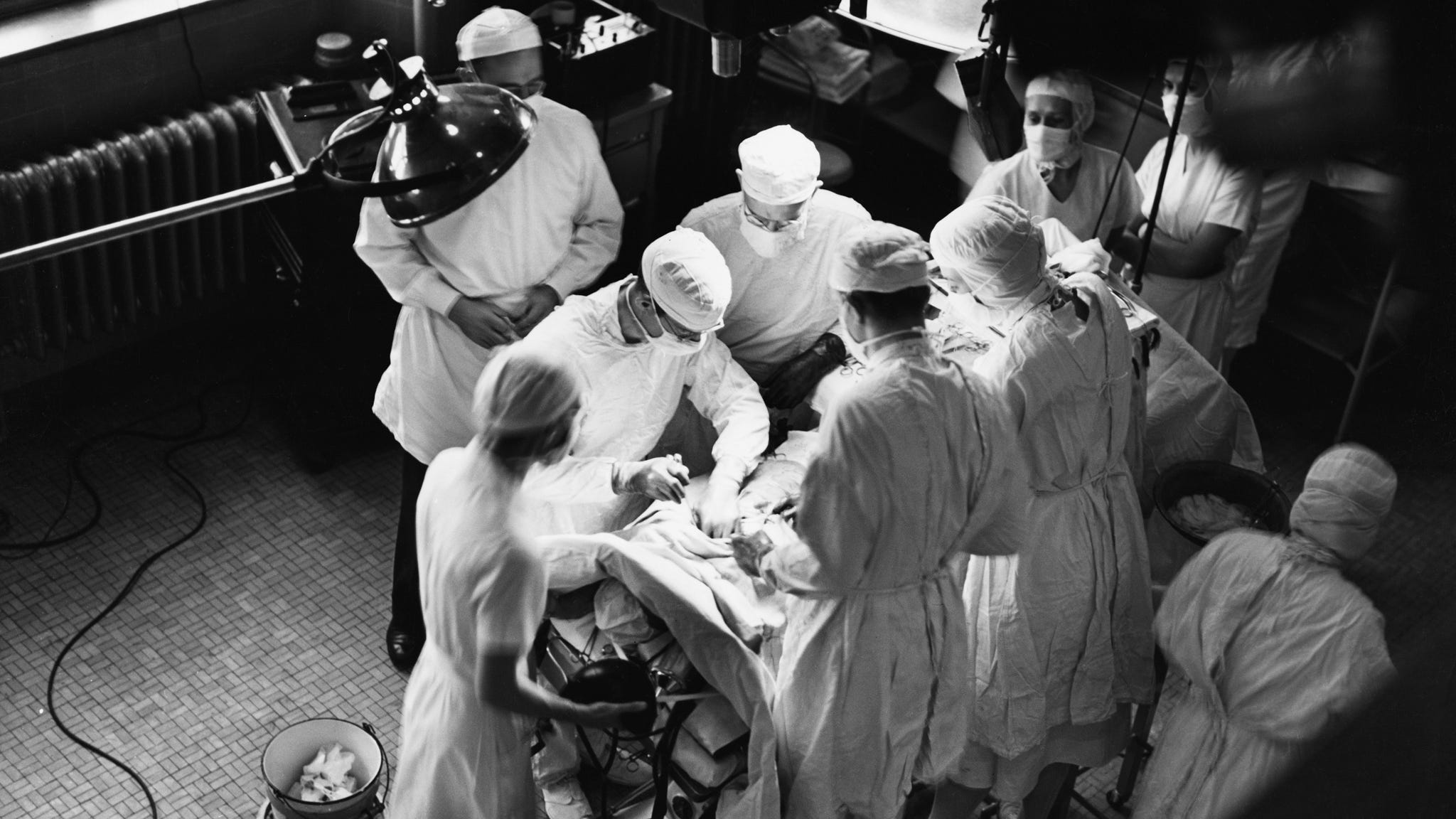
In 1945, a chain-smoking surgeon, a deaf female doctor, and a self-taught African-American lab tech developed a risky procedure that revolutionized medicine.

The Underdog Doctors Who Saved America’s “Blue Babies”

One afternoon in November 1944, the chief of surgery at Johns Hopkins Hospital in Baltimore, Alfred Blalock, sat in his office deep in thought. As usual, he had a cigarette on the go: even after losing two years of his early career to tuberculosis, he had never quite managed to give up his 40-a-day habit. With his neatly combed hair, immaculate chalk-stripe suit and donnish glasses, he might easily have been mistaken for a prosperous lawyer, but at the age of 45, he was already known as one of America’s foremost clinical researchers. A few years earlier he had revolutionized the treatment of circulatory shock, a life-threatening condition in which blood loss makes it difficult for the heart to pump enough fluid to the body. Shock was one of the biggest killers in wartime, frequently the consequence of injury by shrapnel or explosives. Blalock’s experiments led to the routine use of blood-plasma transfusions to treat those with severe wounds, a measure which saved the lives of thousands of servicemen in the Second World War.

This achievement alone was enough to ensure Blalock’s place in the medical pantheon, but this afternoon he felt only frustration. When his senior resident surgeon, William Longmire, walked into the room he found his boss sitting disconsolately behind a pile of books. In recent weeks Blalock had attempted a series of ambitious and difficult procedures on patients seriously ill with abdominal disorders: none had been successful, and most of the patients had died.

“Bill, I am discouraged,” he said to Longmire. “Nothing I do works.”

Blalock was desperate to make an original contribution to surgical history and to silence colleagues who complained that he was a competent researcher but a mediocre clinician. Recently he had been concentrating his efforts on developing new methods to treat problems with the pancreas and intestines. But only a few days later he would perform a novel and entirely different kind of operation, one that would catapult him to fame and make Johns Hopkins a place of pilgrimage for patients and surgeons from all over the world.

Shortly after their conversation, Longmire was summoned to the third floor of the clinic, where Blalock took him to a cot containing one of the hospital’s youngest patients. Her name was Eileen Saxon, and she had been born at Johns Hopkins the previous year. Now 15 months old, she was desperately ill and being kept alive in an oxygen tent. Longmire was shocked by her condition. She was unusually small for her age, but the first thing he noticed was her color. Her skin had a deathly pallor, and her lips and fingernails were a dark, inky blue. Eileen was suffering from a congenital condition called tetralogy of Fallot; children unlucky enough to be born with it were known as “Blue Babies,” and there was little that could be done for them. The blue tinge to Eileen’s skin was cyanosis, the result of blood bypassing the lungs and circulating through the body unoxygenated. Half of all children in her position would die before the age of three, and fewer than a quarter would make it to the age of ten. Those who survived for any length of time endured a miserable existence. Many physicians believed that the smallest degree of excitement would be fatal, and everyday pursuits — school, outdoor play, the cinema, even travel by motor vehicle — were often prohibited. Eileen’s future looked bleak.

When Blalock told his junior that he intended to try a new type of operation on her, Longmire was horrified: given her state, he could not believe that she would survive an anesthetic, let alone a procedure that had never before been attempted. The chief anesthetist, Austin Lamont, concurred. When he heard of Blalock’s plans he flatly refused to take part in proceedings, and the operation was cancelled. But one of Lamont’s colleagues, Merel Harmel, was prepared to take the risk, and it was rescheduled for the following day.

Early on the morning of Wednesday, November 29, little Eileen was taken into room 706, an operating theater on the seventh floor of the building that in later years would be known simply as “the heart room.” Two large windows provided most of the light; in the summer these were usually thrown open in a futile attempt to gain some respite from the fierce Maryland heat. There was a small observation gallery overlooking the operating table, and several hospital staff were leaning over its rail, having heard rumors that something unusual was about to take place. As Blalock scanned the faces of the spectators he caught sight of his laboratory assistant and called out to him: “Vivien, you’d better come down here.”

Vivien Thomas

In his early thirties, Vivien Thomas was a talented surgical technician with a sophisticated grasp of anatomy and physiology — and almost entirely self-taught. Thomas had planned to become a doctor, but his chances of going to university evaporated after the bank holding his savings collapsed in the aftermath of the Wall Street crash. Instead, he found a job working in Blalock’s laboratory in Nashville, where the authorities paid him a pittance and categorized him as a janitor because he was black. He soon became so essential to Blalock’s work that when the surgeon moved to Baltimore he insisted that Johns Hopkins employ Thomas too. Since he had no medical qualifications, Thomas usually had no contact with patients. His role was in the animal laboratory, where he performed physiological experiments and helped to develop new surgical procedures. He had perfected the operation Blalock was about to perform, practicing it hundreds of times on dogs and refining every detail.

But the idea to operate on these desperately ill children had come from one of the other people standing expectantly in the operating theatre. Helen Taussig was the pediatric cardiologist looking after Eileen Saxon; like Thomas, she had faced prejudice and personal setbacks in her early career. As a student, she had been rejected by Harvard Medical School on the grounds that she was a woman, but had nevertheless managed to become the world’s leading expert on congenital heart conditions. Profoundly deaf since her thirties, she had taught herself to diagnose rare conditions using her hands as a stethoscope.

Thomas and Taussig would play no active part in proceedings, but Blalock leaned heavily on them for advice. The surgical team assisting him was young and formidable; it included William Longmire and William Muller, two outstanding protégés who would soon be appointed to major professorships. Standing at the foot of the table, ready to administer intravenous fluids to the patient, was a 24-year-old intern whose career would outshine even theirs: his name was Denton Cooley, and he was destined to become one of the most celebrated surgeons in the world, and the first to implant an artificial heart.

The mood was far from optimistic. Longmire was convinced the girl would die, and Cooley’s considered opinion was that the operation would be “a big disaster.” Merel Harmel put a mask over Eileen’s face and dripped ether on to it. This was a primitive way of putting a patient to sleep, used since the dawn of anesthesia in the 1840s; it was also dangerous, as it was difficult to control how deeply or for how long the patient would remain unconscious. As Eileen succumbed to the ether, Blalock and his colleagues — eight in total — gathered around her tiny body. Vivien Thomas remarked that she was so small that it was difficult to believe that there was a patient underneath the sterile drapes.

Blalock made the first incision on the left side of her chest, starting at her breastbone and extending it to her armpit. As he cut through her muscle in order to gain access to the heart, blood welled up from a number of small arteries. The surgeons were taken aback by its appearance: rather than the free-flowing, bright red fluid they were used to seeing, this was a glutinous blue-black. Longmire described it as “like purple molasses”; had he been in charge, the operation would have been abandoned there and then.

The alarming color of Eileen’s blood was the cause of her infirmity. Although people often refer to “the circulation” when talking about the movement of blood, there are really two: the pulmonary circulation and the systemic. When the blood has completed its journey around the body and arrives back at the heart it has given up most of its oxygen to the organs and tissues; in this state, it has a blueish tint. The right side of the heart then pumps it to the lungs, where it passes through tiny vessels that allow freshly inhaled oxygen to pass into the red blood cells. At the same time, carbon dioxide — a waste product of processes inside the body’s cells — moves in the opposite direction and is then exhaled. The freshly oxygenated blood, now bright red, travels back to the left side of the heart, ready to be pumped through the rest of the body. Blood, therefore, passes through the heart twice: first as part of the pulmonary circulation through the lungs, and then as part of the systemic circulation that nourishes all our major organs.

In tetralogy of Fallot, this neat arrangement is hopelessly compromised. Those with the condition have four separate cardiac deformities, of which two are responsible for the characteristic skin pallor. Whereas the left and right sides of a normal heart are separated by a wall of tissue, in tetralogy there is a large aperture between the two, known as a septal defect. As a result, oxygenated blood from the lungs mixes freely with blue, deoxygenated blood from the rest of the body. In addition, the pulmonary artery, the vessel through which blood is pumped to the lungs, is drastically narrowed so that its flow is greatly reduced. When the heart of a tetralogy patient contracts, only a small proportion of the deoxygenated blood in the right ventricle is able to escape through the narrowed pulmonary artery towards the lungs; most of it instead passes through the septal defect and into the systemic circulation. Because so little of their blood has travelled through the lungs, Blue Babies have extremely low blood oxygen levels, causing breathlessness, stunted growth and the unhealthy coloration which typifies the condition.

Blalock could not entirely correct this malformation, but by some ingenious plumbing, he hoped to improve Eileen’s condition. Having exposed her heart and its major vessels, his plan was to redirect one of her arteries so that instead of delivering blood to her left arm it would instead send it back toward her lungs, increasing her overall oxygen levels. In surgical parlance this type of procedure, in which blood is redirected to where it is most needed, is called a “shunt”; within a few years Blalock’s operation would be universally known as the “Blalock-Taussig shunt.” (It has since been argued, with some justification, that given the importance of Vivien Thomas’s contribution the operation should instead be known as the Blalock-Taussig-Thomas shunt.)

The first challenge was identifying the correct blood vessels. Unless you have seen the inside of the human body first hand, it’s difficult to appreciate how little it resembles the neat arrangement of nerves and vessels depicted in a textbook. Every patient is different: arteries vary greatly in size or trace entirely unexpected paths; blood vessels sit so close together that they become almost impossible to differentiate. And they lie snugly embedded in tissue, requiring meticulous dissection in order to lay eyes on them in the first place. It took Blalock some time to be sure he had found the two arteries he was looking for.

The first of these was the left pulmonary artery, which takes blood from the heart to the left lung. The procedure would involve shutting off this vessel for as much as half an hour, with the result that Eileen would be breathing through a single lung. This was a huge risk in an already oxygen-deprived patient, and so Blalock temporarily clamped the vessel to see how she would tolerate it. To everybody’s alarm, her skin became an even more icy blue, as if her life was ebbing away before their eyes. Harmel attempted to put a tube into the girl’s windpipe in order to deliver oxygen straight to her one functioning lung, but without success: endotracheal tubes for such small children had not yet been manufactured, and the only implement available — a urinary catheter — was hopelessly unsuited to the job.

There was an anxious wait as the surgeons decided what to do. To their relief, Eileen’s color improved slightly without further interference; they could continue. With infinite care, Blalock freed a second artery from its surrounding tissue. This was new territory for him: although he had watched Vivien Thomas carry out the procedure on dogs, he had never attempted it himself. He had installed Thomas — the expert, with 200 trial runs under his belt — on a stool behind him, a vantage point from which he could observe and make suggestions. Periodically Blalock would turn to his assistant and ask for advice: is this artery long enough? Is this the right part of the vessel to clamp?

Eileen’s blood vessels were even smaller than those of the dogs Thomas had operated on: the one Blalock needed was no bigger than a matchstick. This was the vessel taking blood to her left arm, the left subclavian artery. After isolating a section of the vessel with clamps to ensure no blood was flowing through it, he cut through the artery with a scalpel. He made a small incision in the side of the pulmonary artery and began the most challenging part of the procedure. A fine needle threaded with silk was used to attach the tiny subclavian artery to the pulmonary artery. Placing the sutures required almost inhuman accuracy, and the instruments at Blalock’s disposal were ill-suited to such delicate work. Nothing of this kind had been attempted before on the miniature vessels of a child: the needle and forceps Blalock was using were designed for adult surgery and felt unpleasantly cumbersome as if he were trying to repair a Swiss watch using a plumber’s wrench.

After what seemed like an eternity, the two vessels were united. In theory, a proportion of the blood leaving the heart would be pumped into the subclavian artery and then be redirected back into Eileen’s left lung, increasing its oxygenation. But the vessel was so small that Blalock was unsure if the operation would have much effect. He released the clamps, allowing blood to pass through the new junction for the first time. After checking carefully for signs of bleeding, he closed the chest and stitched the external wound. The operation had lasted a little over an hour and a half; although there was no obvious improvement in Eileen’s condition, she had survived.

Complications followed, however, and for two weeks the little girl’s life hung in the balance. Thereafter she began to make good progress, and on January 25, almost two months after the operation, she was well enough to go home. Helen Taussig was pleasantly surprised to find that she had started to gain weight, and her episodes of cyanosis became less pronounced. Her parents were delighted: previously too ill to go outside, she was now learning to walk and could join them on outings to their local park. The operation had been a modest but definite success.

Despite this initial improvement, Eileen Saxon lived only a few months longer: her symptoms returned and a second operation was unsuccessful. It later became clear that children under the age of three gained only temporary benefit from the procedure.

This outcome seemed to justify further attempts, and on February 3, Blalock operated on a second patient, this time a nine-year-old girl. On her arrival at the hospital she was only able to walk 30 feet, stooping and panting; a month after surgery she could walk upright for twice that distance, without any sign of discomfort. This was an encouraging development — but better was to come. On February 7, 1945, a desperately ill six-year-old boy was admitted to Johns Hopkins. He was extremely undernourished and could manage only a few paces without losing his breath. Taussig examined him and noted that he was severely cyanosed, recording in her notes that the insides of his cheeks were a deep mulberry color. His parents were adamant that they would take any chance to save their son, and three days later Blalock operated.

This time he used the innominate artery, a vessel supplying blood to the arm, head and neck. First, he severed it just before the point at which it split into the two branches supplying the head and left arm. After suturing its upper extremity closed, he attached the lower part of the vessel, the end nearer the heart, to the right pulmonary artery. This new circuit would redirect some of the boy’s blueish systemic blood back into the lungs for an additional dose of oxygen. When he released the clamps, a stream of blood gushed out of an undetected hole between the sutures; he quickly reapplied the clamps to cut this off and repaired the opening with an extra stitch. This time the join was perfect, and when the circulation was allowed to flow again it did so without any problems.

As the patient’s right lung received its first blood in over an hour, something extraordinary happened. Merel Harmel, the anesthetist, suddenly cried, “He’s a lovely color now! Take a look!” Blalock and Taussig moved to the head of the table and were astonished to see a pink-faced little boy with healthy red lips. Within a few minutes, he was awake and asking to get out of bed. Elation filled the room. A young medical student who had been observing the operation, Mary Allen Engle, was so moved that she decided then and there to become a cardiologist; she would go on to be one of the field’s leading authorities. For the next few days, nurses accustomed to tending critically ill children found themselves dealing with a patient who was desperate to run around and play. The transformation was miraculous: in her case notes, Helen Taussig recorded that “his disposition has changed from that of a miserable whining child to a happy smiling boy.”

That dramatic color change — from sickly blue to healthy pink — was a proof of success that both medics and the public could readily understand. Blalock and Taussig wrote a report of their three cases for the Journal of the American Medical Association. Before the article had even been printed a journalist from the Associated Press somehow got hold of a copy, realized its significance, and published a story. Editors leapt upon it: at a time when newspapers were full of harrowing details of American deaths in the war in Europe, this was a rare morsel of good news: a happy story of terminally ill children being magically restored to life.

The effect was electrifying, and the medical world was utterly unprepared for it. Tetralogy of Fallot is one of the more common congenital heart conditions, affecting one in every 2,400 newborns. There were thousands of children in the U.S. alone who might be saved by the new operation, invalids presumed to be beyond medical help. Family doctors who knew nothing about Blalock’s breakthrough suddenly received visits from parents demanding a referral to the hospital in Baltimore. Children began to arrive at Johns Hopkins from across the country and then from abroad, and before long a trickle had become a flood. Not all parents could afford the operation, and some small-town newspapers launched public appeals to fund them. Such patients often turned up with a local journalist in tow, and surgeons found themselves constantly answering questions from uninvited members of the press.

Within a few months, the pediatric beds of Johns Hopkins were full, with tetralogy patients (known to the staff as “tets”) spilling out into the adult wards. This was an exhausting period for Blalock’s team, who were constantly operating during the day and often called to the wards at night; they barely took a day off. Regular delegations of surgeons arrived from abroad to observe the operation and find out what equipment they would need in order to do it themselves.

A still more rapturous reception awaited Blalock in Europe. He was invited to spend a month at Guy’s Hospital in London, and on August 22, 1947, Blalock and his wife Mary sailed for England on the Mauretania. He had spent the day before his departure frantically trying to procure a month’s supply of Viceroy cigarettes, which were unobtainable in Britain. During his short residency at Guy’s, Blalock operated successfully on ten blue babies. His visit was widely reported: one woman from Sevenoaks in Kent, a Mrs. Gallard, read about the celebrated American heart surgeon and promptly jumped on a London train to ask him to operate on her eight-year-old son Roger, who was confined to a wheelchair. Four months later a local newspaper reported that he was now able to run around and play with his friends. His mother explained to the journalist, not entirely accurately, that the surgeon had “removed Roger’s heart, and while it lay pulsating in his hands, he remodeled it.” Displaying a similarly hazy grasp of surgical minutiae, a Daily Mail article the following month claimed that during the procedure another patient’s heart was “removed and massaged.”

Toward the end of his stay, Blalock shared the stage with Helen Taussig for a joint lecture to a packed hall at the British Medical Association. Their presentation ended in spectacular fashion when a spotlight beam suddenly pierced the darkened hall to pick out a nurse sitting on a chair with an angelic and healthy two-year-old on her lap; Blalock had operated on the little girl a week earlier. The leading British surgeon of the day, Russell Brock, described it as “a Madonnalike tableau, a perfect climax to an impressive lecture on an epochmaking contribution.”

Alfred Blalock (center) performs a Blue Baby operation at Johns Hopkins Hospital in Baltimore. Vivien Thomas is standing behind him. (Photo courtesy of Alan Mason Chesney Medical Archives.)

Blalock’s tour (his “royal progress,” as one of his juniors described it) continued with visits to hospitals in Sweden and France. He left behind an important legacy in Europe: a “Blue Baby clinic” was set up at Guy’s, and within a few months the operation was being successfully performed in many other centers across the continent. As they boarded their plane back to America, Blalock remarked to his wife that they had been treated like gods; now they must come down to earth again. This was no exaggeration: a massive backlog of cases awaited him in Baltimore. By 1948 there was a three-year waiting list for the operation, and two years later a photographic portrait of Blalock was commissioned to commemorate the 1,000th case.

Despite the hundreds of lives it saved, the work of Blalock, Taussig and Thomas was also fiercely criticized. Antivivisection campaigners were outraged at the number of animal experiments taking place in the Johns Hopkins laboratories and fought tirelessly to put a stop to them. Experimental surgery on animals had long been standard practice in developing new operations; dogs, whose hearts and major vessels were similar in size to those of humans, were easily available since most cities had large numbers of unwanted strays. But some of these experiments were of dubious value and inflicted unnecessary suffering. In 1901 one of the pioneers of cardiac surgery, Benjamin Merrill Ricketts, performed experiments on 45 dogs at his laboratory in Cincinnati, deliberately injuring their hearts and surrounding tissues. “The object was to induce as many complications as possible,” he explained, and in this, he was certainly successful: the vast majority of the animals died shortly afterward.

By the 1940s researchers had a more enlightened attitude, and Vivien Thomas’s surgical trials on dogs were all conducted with full anesthesia; those that developed complications were humanely killed. Nevertheless, there were repeated attempts to shut down his research, with campaigners harassing laboratory staff and the suppliers of animals. A national animal-rights movement had emerged in the 1880s and later succeeded in presenting to Congress a bill to ban animal experimentation. It was defeated, but vigorous local campaigns continued in many states. In February 1946, as Congress considered new antivivisection legislation, Blalock gave evidence before a House committee with three of his young patients, explaining that without animal experimentation none of them would have survived. He made a powerful case, and the bill was voted down.

Local activists in Baltimore were more tenacious and succeeded in preventing medical researchers from using unclaimed stray dogs for their experiments. Without a ready supply of animals, hospitals took to buying them in from neighboring states, and when this practice was also deemed unlawful matters came to a head. Baltimore City Council announced a public vote — popularly known as the “dog referendum” — to decide whether the use of animals for medical research should be prohibited. The pro-vivisection camp indulged in some emotive tactics in its attempt to win the public over. When Helen Taussig spoke at an open meeting she was accompanied by a brigade of her patients: healthy, smiling children, many of whom had brought their pet dogs. The star of the campaign was a playful and photogenic mongrel called Anna, an early survivor of Vivien Thomas’s surgical trials. She appeared in an educational film and was photographed for Life magazine with one of the children who had been saved by the operation. The result of the dog referendum was decisive: the antivivisection faction was resoundingly defeated by a margin of more than four to one. To commemorate the win, Blalock commissioned a portrait of Anna, which remains on display at the hospital today.

Blalock and his first Blue Baby success of 1944 captured the public attention as no operation had before. Most modern surgeons would agree with Russell Brock’s assessment that it represented the starting point of modern heart surgery, although Blalock had not operated on the organ itself, but on the blood vessels around it. Nor had he cured his patients: the operation was palliative, improving their quality of life rather than correcting the underlying condition. It was not until a decade later that another surgeon, C. Walton Lillehei, succeeded in curing a cyanotic infant.

Tetralogy of Fallot is only one of a vast array of cardiac malformations, many of which at the time offered a still gloomier outlook for patients, with no hope of a cure. The battle against congenital disease was only just beginning, but Blalock had made an effective assault on its ramparts.

* * *

This story is excerpted from The Matter of the Heart: A History of the Heart in Eleven Operations, now available from St. Martin’s Press.